Retinal DiseaseDeployed
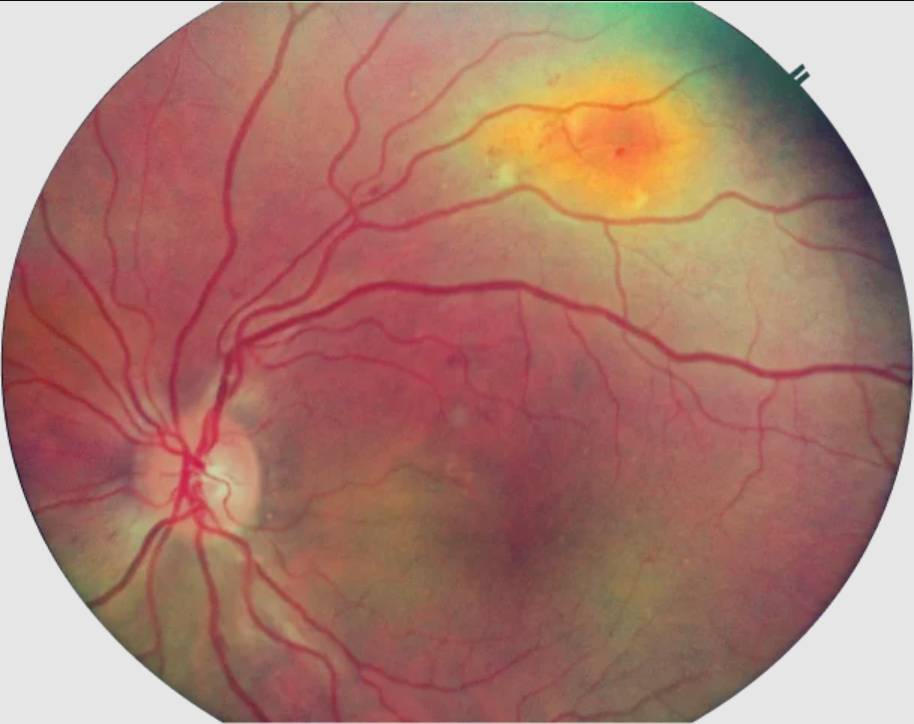
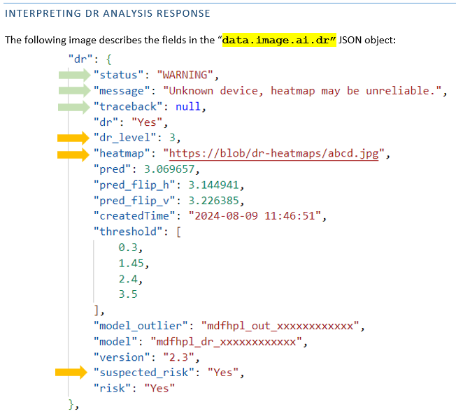
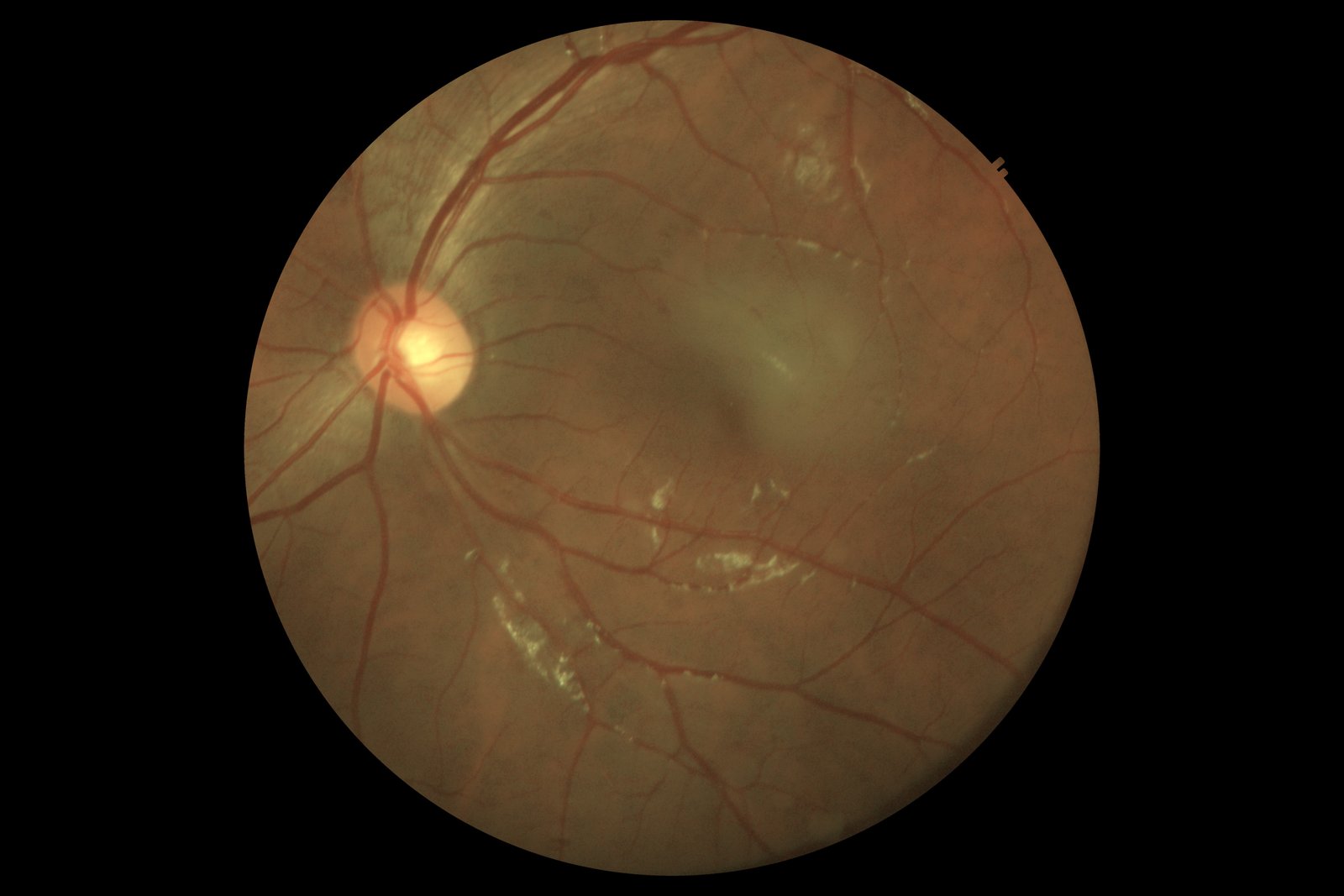
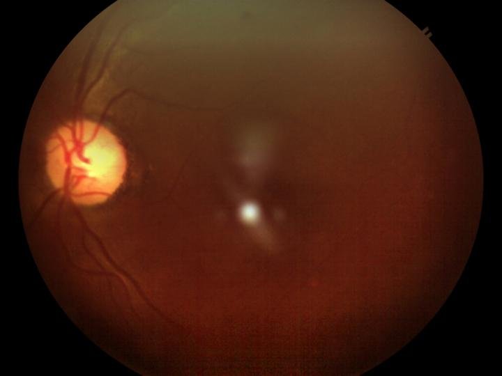
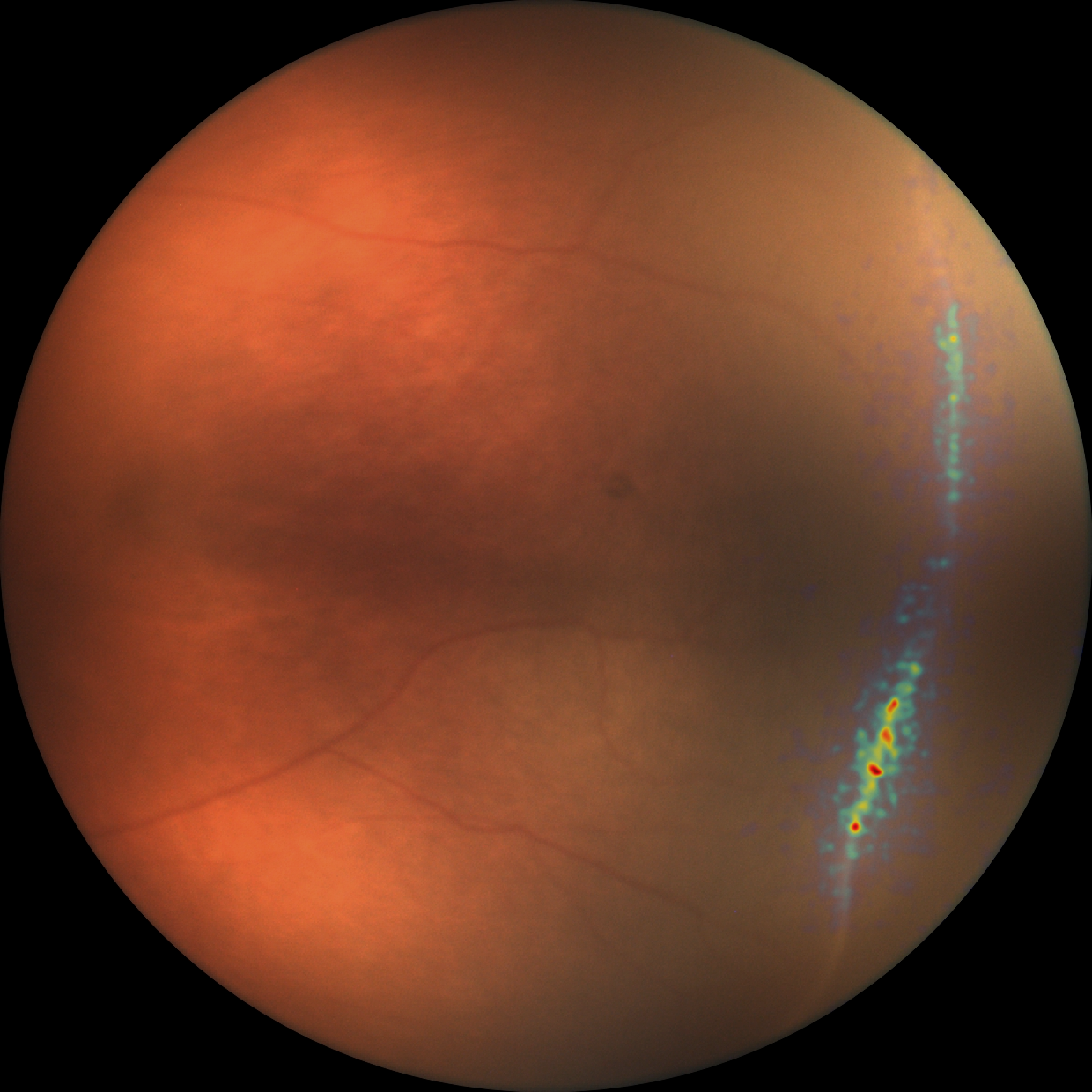
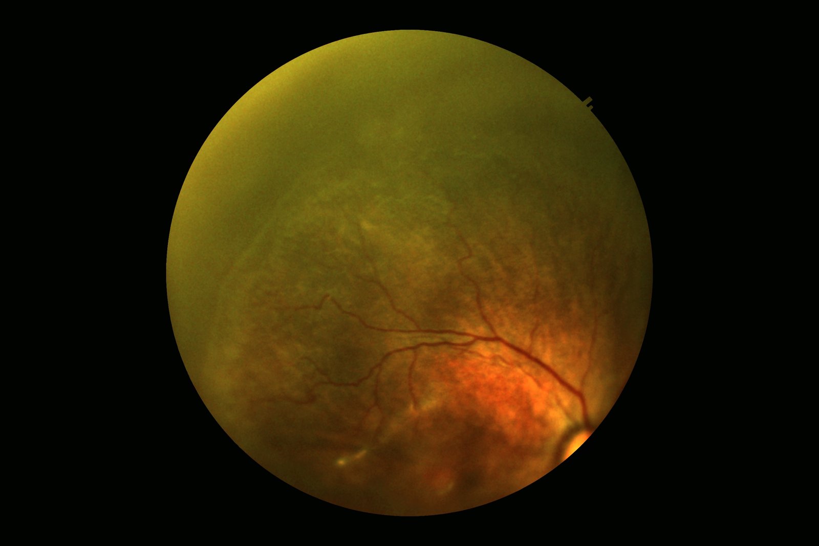
Diabetic Retinopathy Grading ★ Flagship
Deep CNN classifier trained on 500K+ real-world screenings. Detects microaneurysms, hemorrhages, exudates,
and neovascularization. Produces No DR → Proliferative scores with Grad-CAM heatmaps and macular edema
co-detection.
EfficientNet-B4Multi-class
classificationGrad-CAMMacular edema
co-detection
Retinal DiseaseDeployed
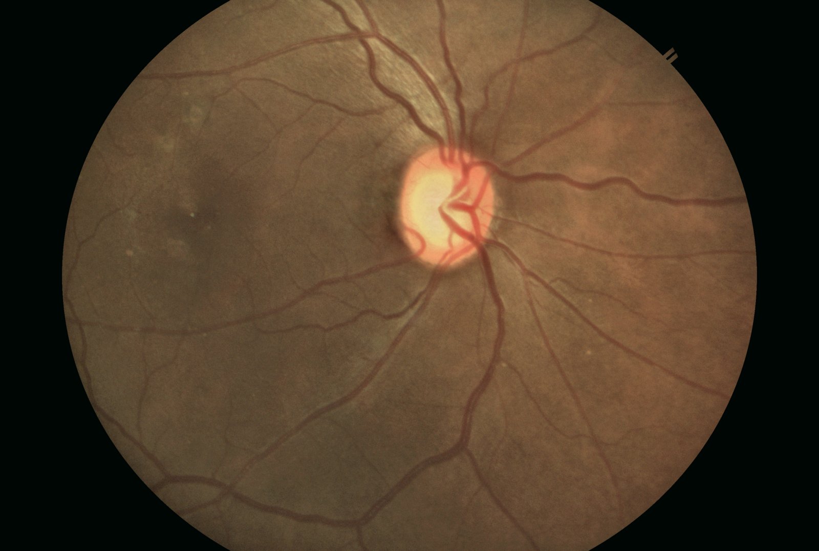
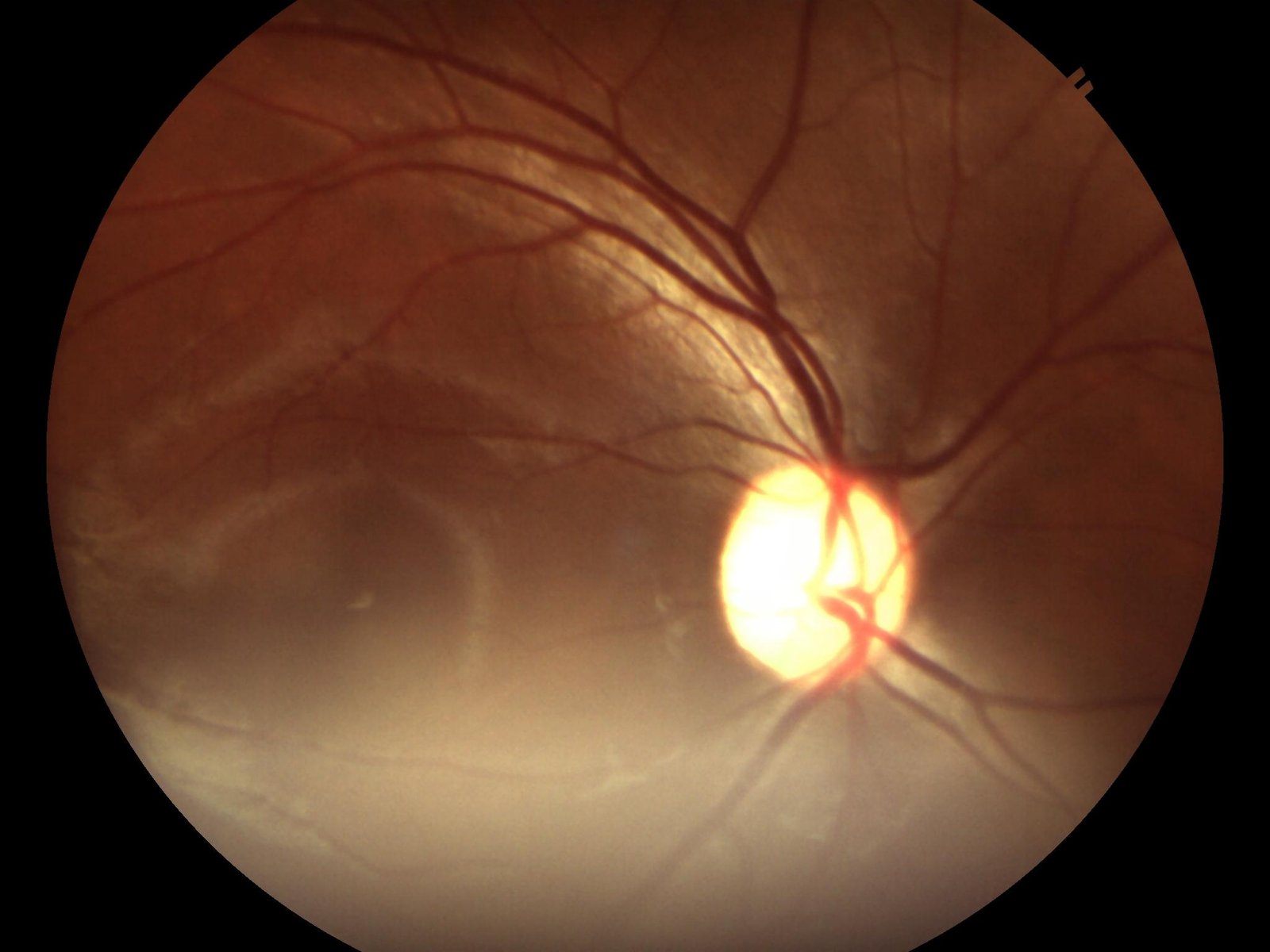
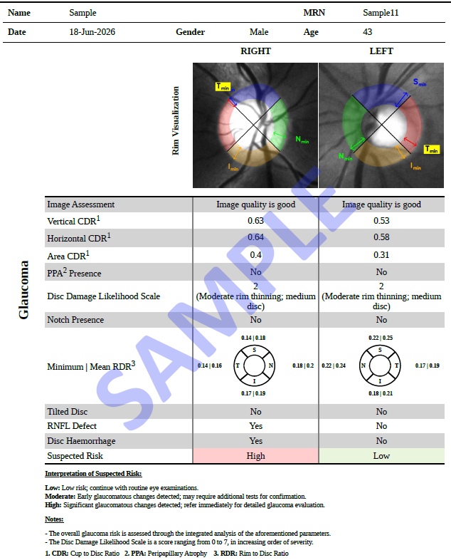
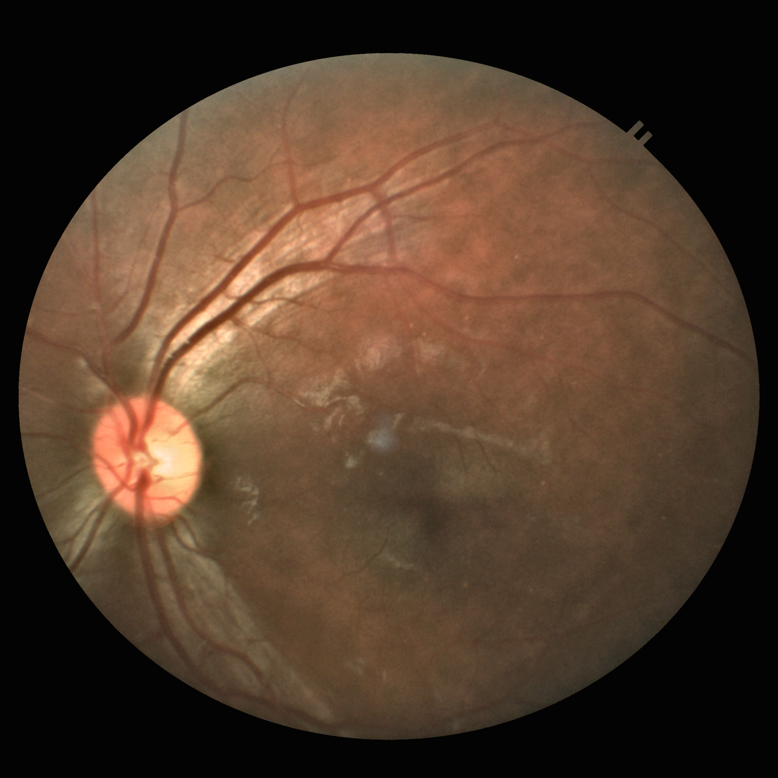
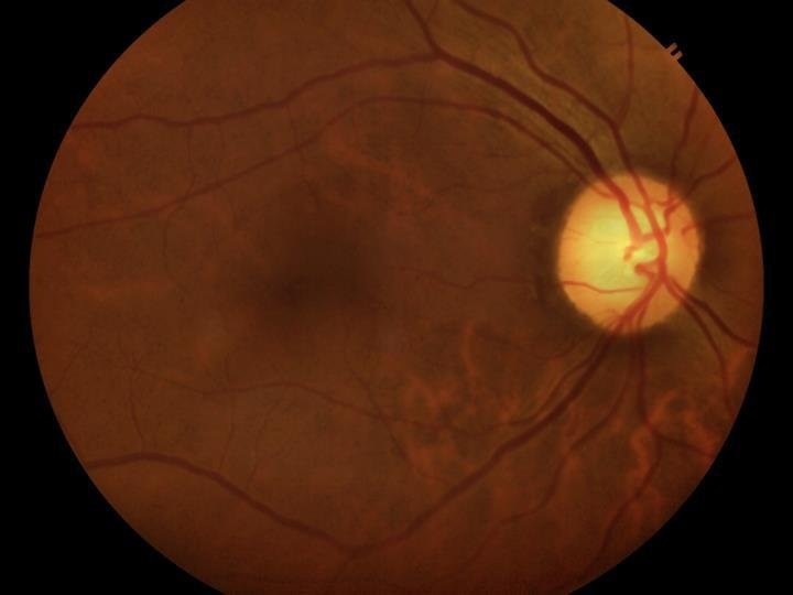
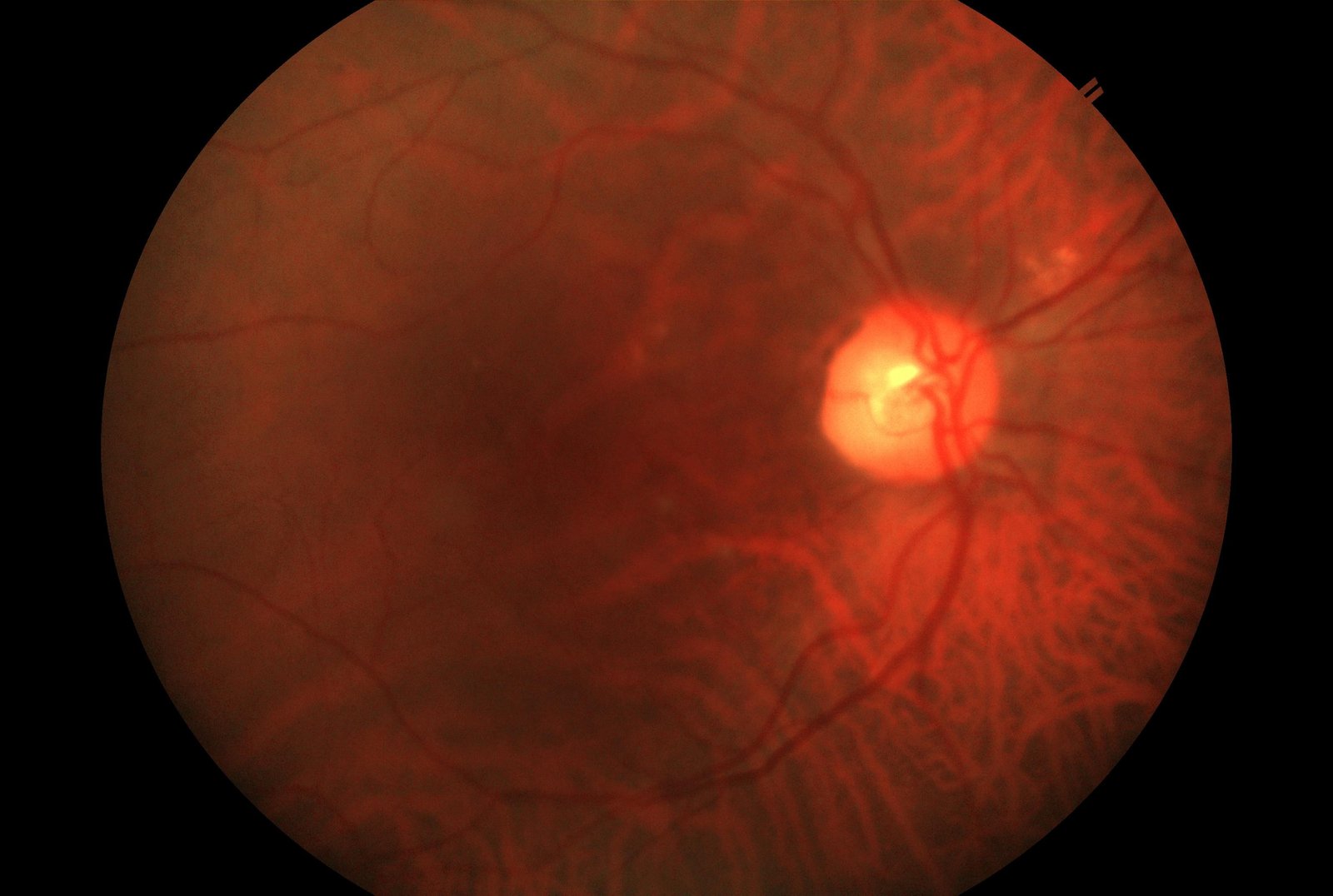
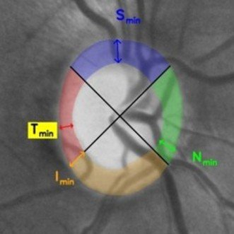
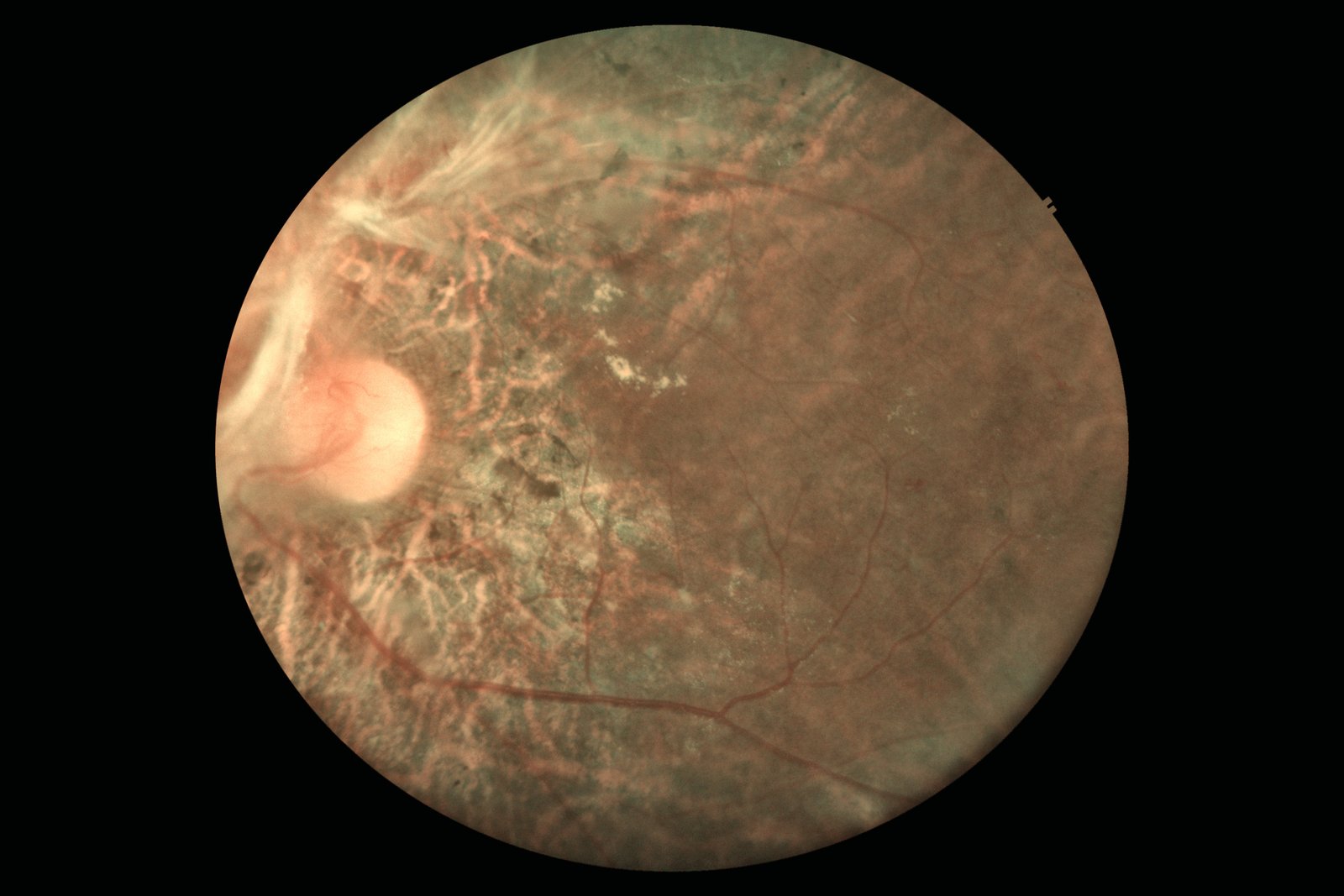
Glaucoma Suspect Detection
Automated optic disc and cup segmentation with cup-to-disc ratio estimation. Identifies RNFL thinning and
disc pallor indicative of glaucomatous damage. Outputs suspicion flag with segmentation overlay for referral
triage.
Attention U-NetSegmentation +
ClassificationCDR estimation
Retinal DiseaseDeployed
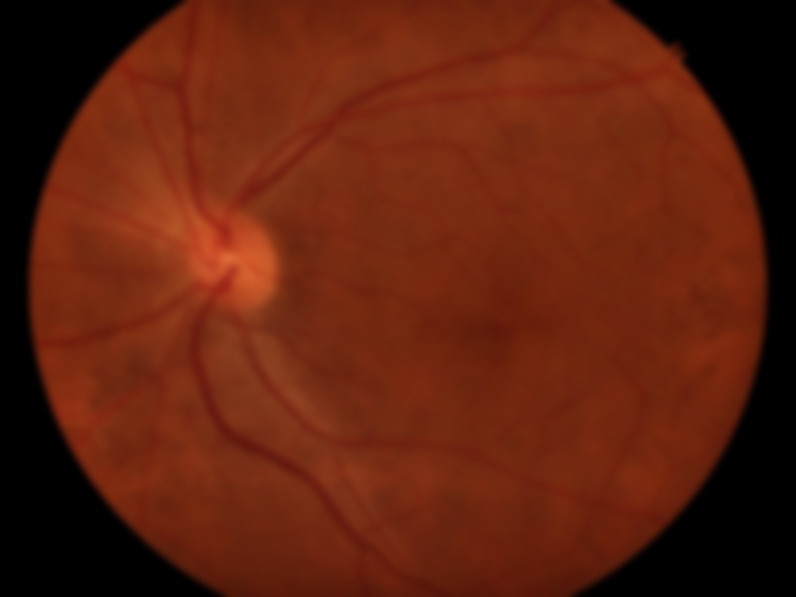
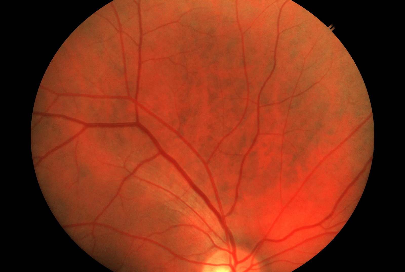
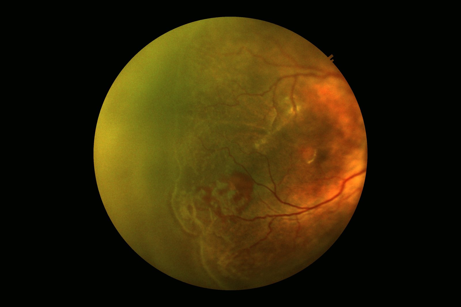
Age-Related Macular Degeneration
Detects early, intermediate, and late AMD by identifying drusen deposits, geographic atrophy, and choroidal
neovascularization. Supports dry vs. wet AMD differentiation to guide urgency of referral.
EfficientNet-V2Drusen segmentationAREDS grading
Retinal DiseaseDeployed
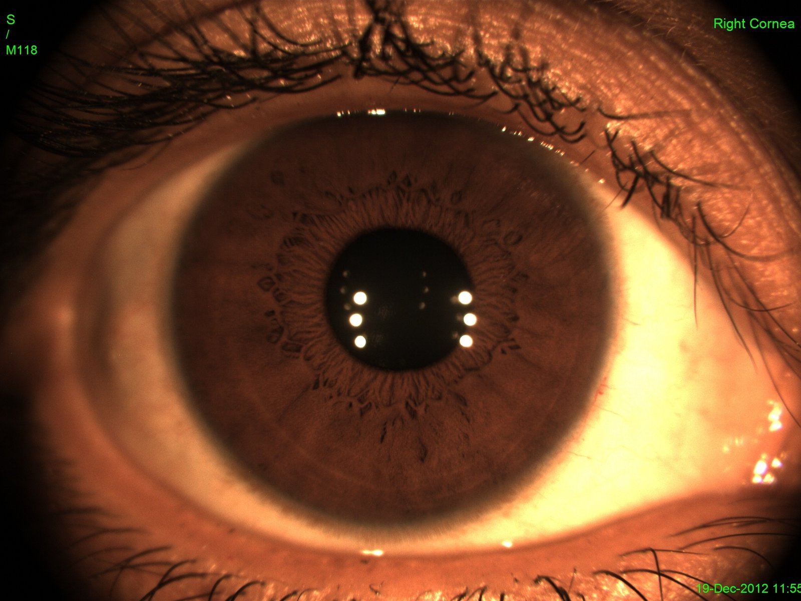
Cataract Detection & Grading
Detects lens opacity and grades cataract severity from retinal images using image quality degradation as
proxy signal. Enables opportunistic cataract screening during routine retinal examination — no separate
slit-lamp required.
Image quality regressionLens opacity
scoringReferral calibration
Systemic BiomarkerDeployed
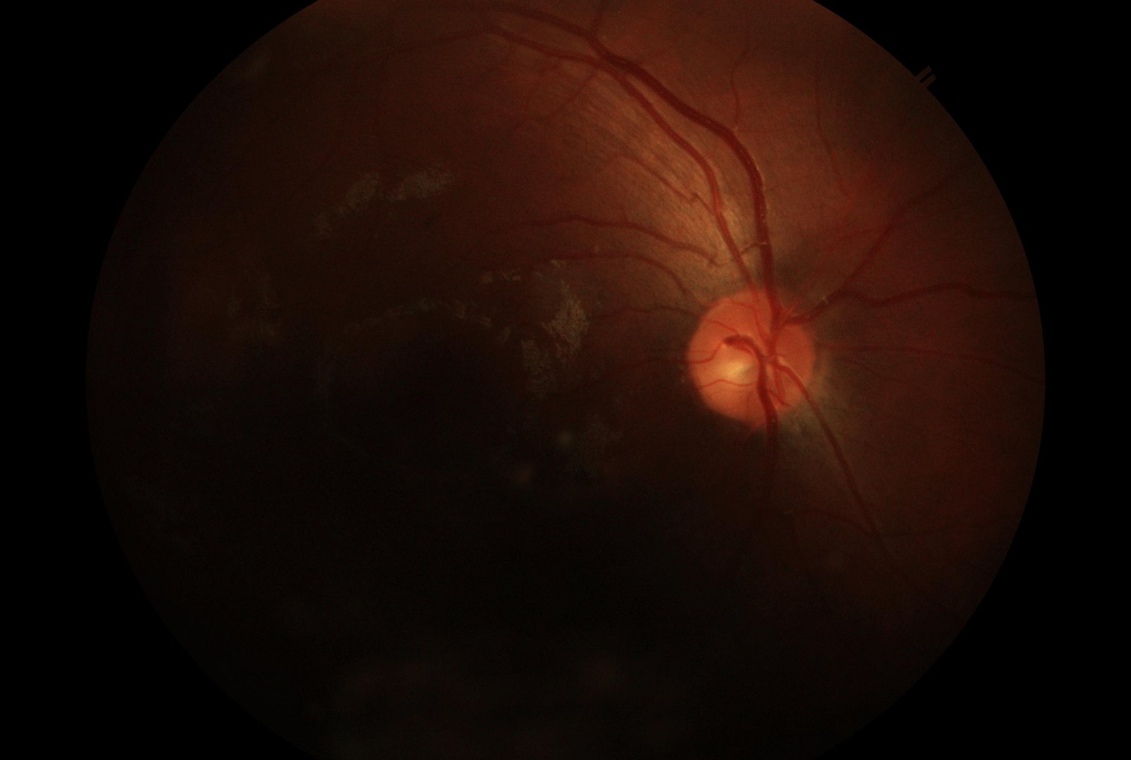
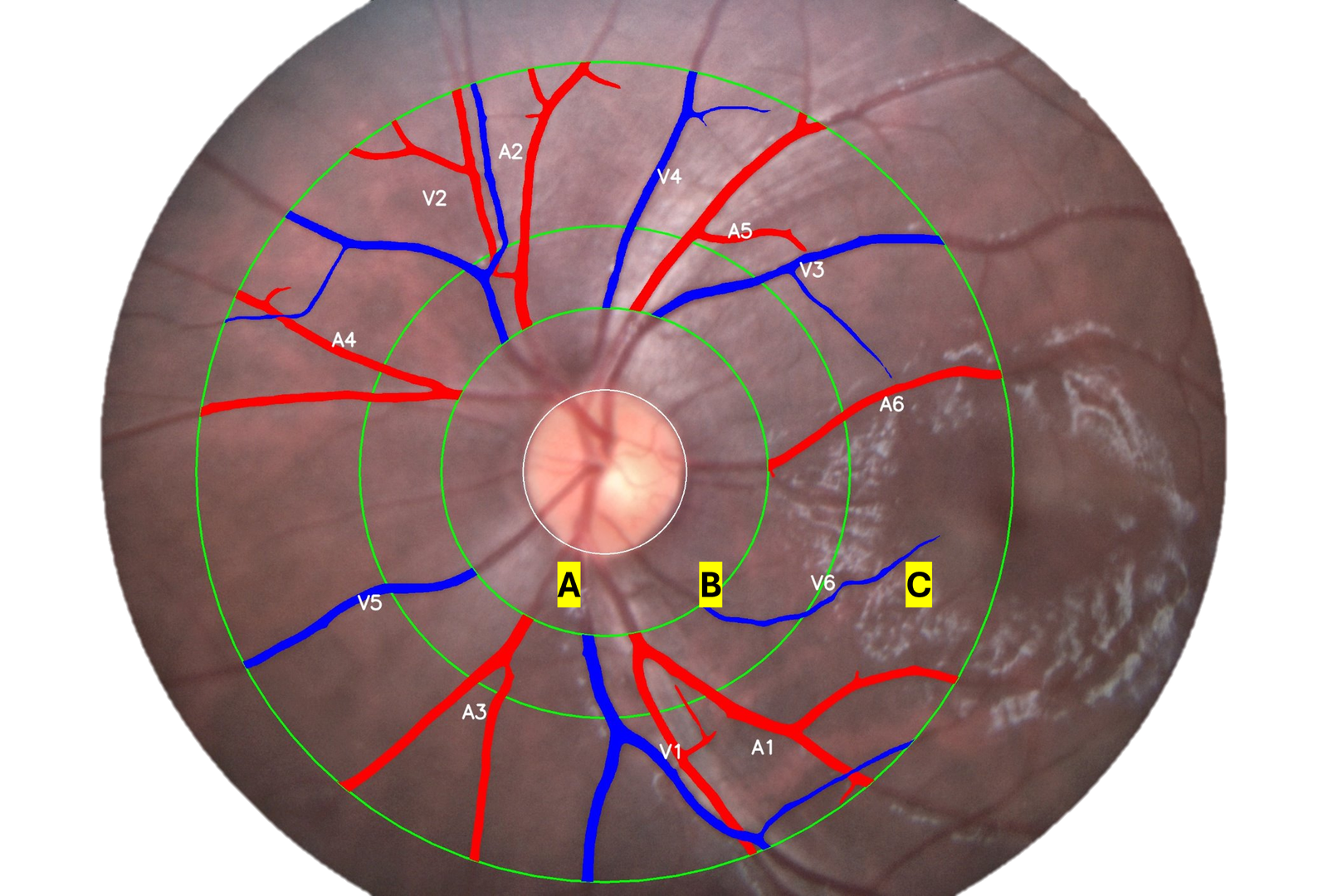
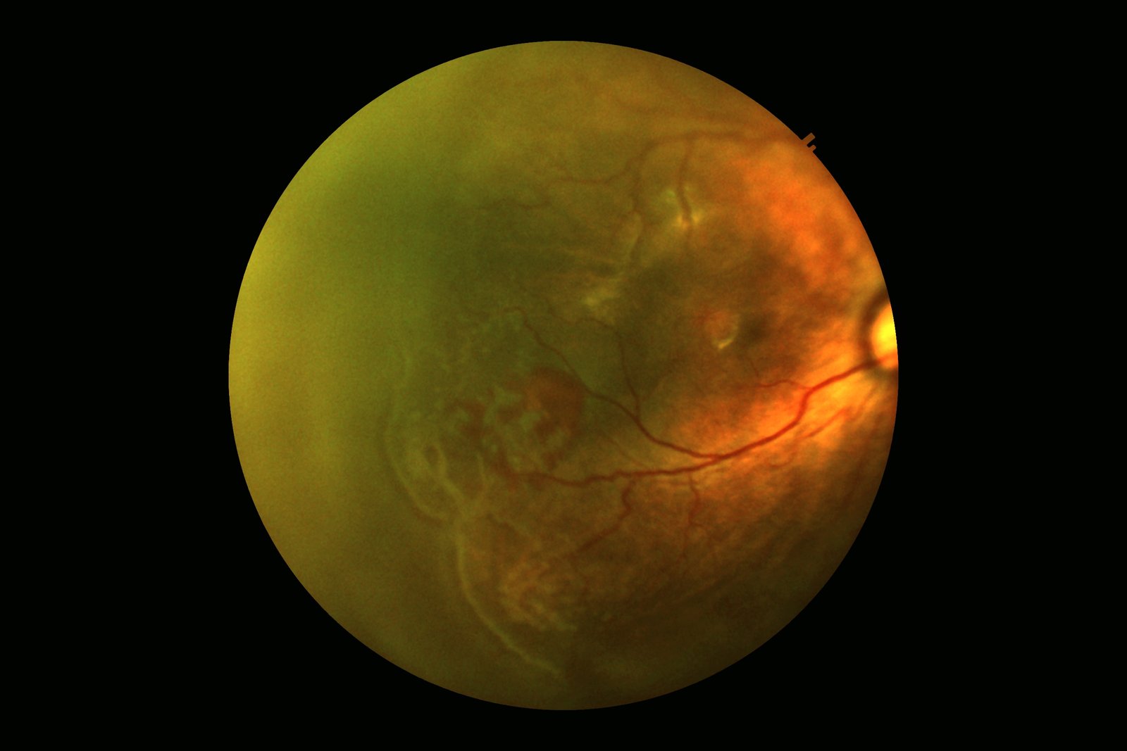
Hypertensive Retinopathy
Identifies vascular changes associated with systemic hypertension — arteriovenous nicking, silver/copper
wiring, focal arteriolar narrowing. Non-invasive hypertension screening signal for community health
programs.
ResNet-50Vascular feature
extractionA:V ratio estimation
Systemic BiomarkerIn Validation
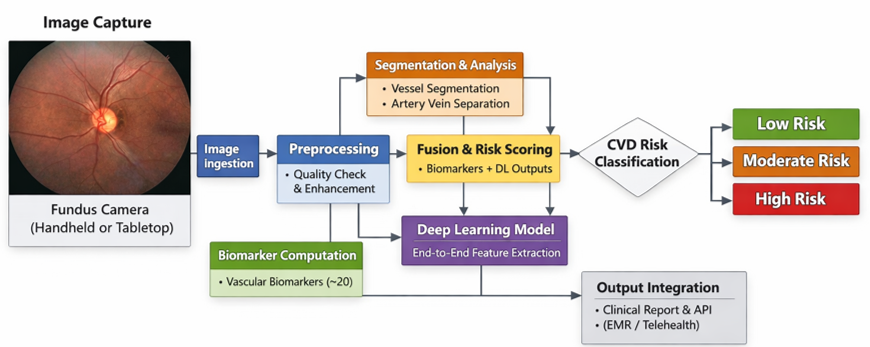
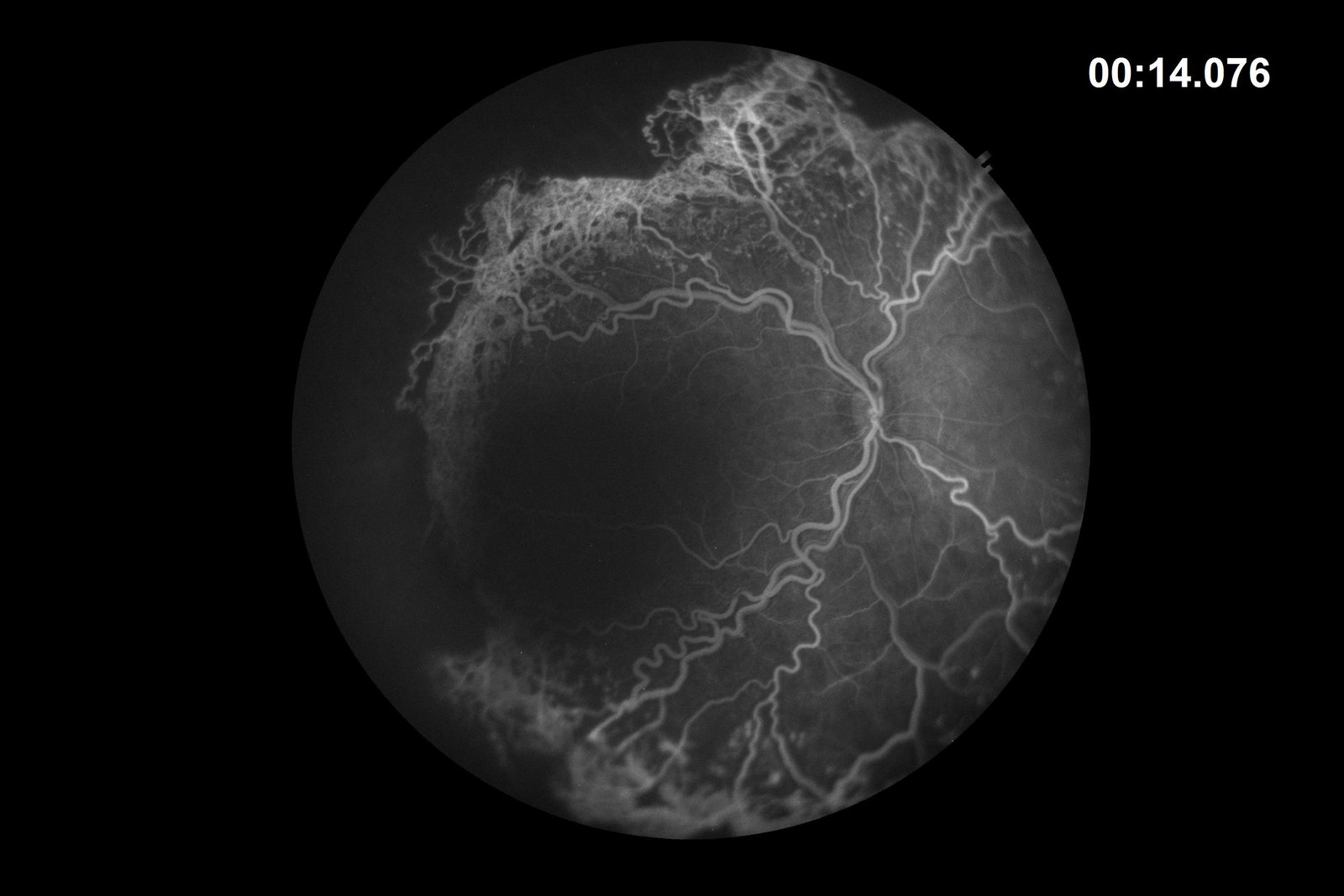
Cardiovascular Risk from Retina
Leverages retinal vascular geometry — fractal dimension, vessel tortuosity, caliber asymmetry — as
surrogate markers for CVD risk. Predicts composite cardiovascular risk score from fundus image alone,
without blood tests or ECG.
Vision Transformer (ViT)Vascular
geometryMulti-ethnic validation
Anterior SegmentDeployed
Lipid Layer Interferometry (LLI)
Analyzes interference fringe patterns in the tear film lipid layer. Classifies lipid layer thickness and
uniformity as an objective dry eye biomarker — replacing subjective grading with AI-scored metrics
correlated with OSDI severity.
Texture CNNFringe pattern analysisOrdinal classification
Anterior SegmentDeployed
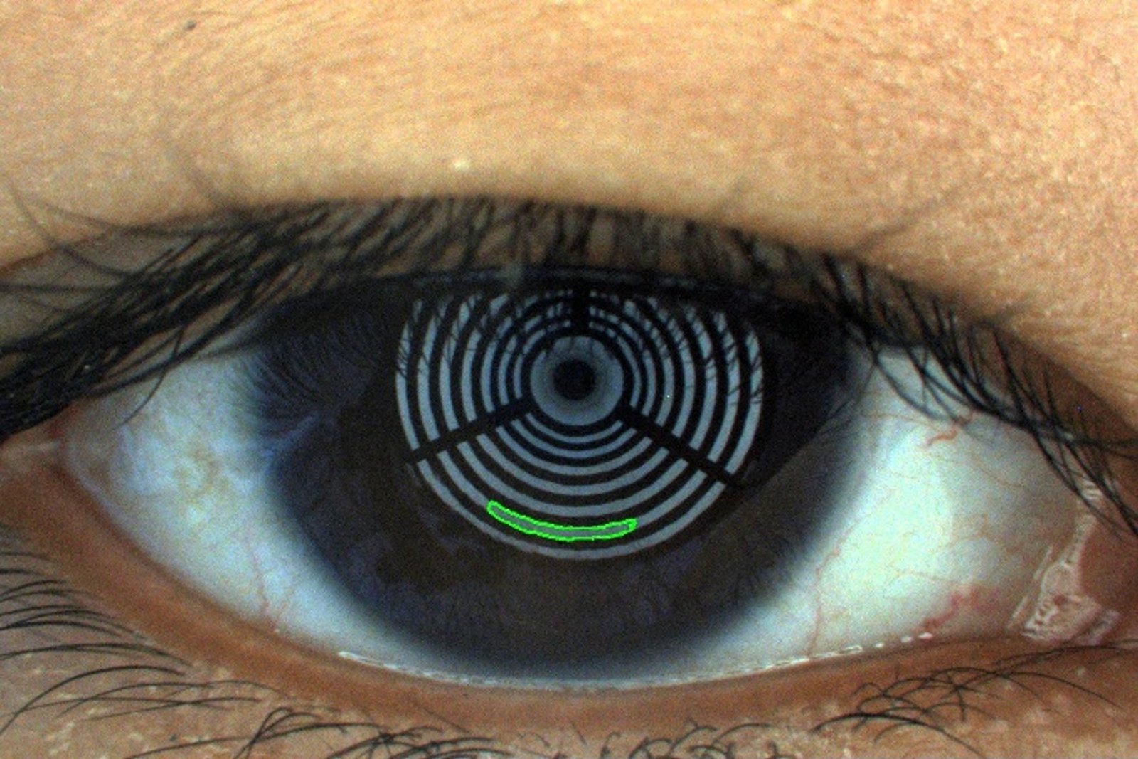
Tear Meniscus Height (TMH)
Sub-mm
Measurement precision
Automated measurement of tear meniscus height from anterior segment slit images using semantic
segmentation. Provides an objective tear volume surrogate — a key parameter in the TFOS DEWS II dry eye
diagnostic algorithm.
Semantic segmentationSub-pixel
regressionDEWS II integration
Anterior SegmentDeployed
Meibomian Gland Dysfunction (MGD)
Segments and quantifies Meibomian gland morphology from infrared meibography. Computes gland dropout score,
tortuosity index, and gland density — fully automated pipeline replacing manual grader assessment.
Instance segmentationGland dropout
scoringTortuosity index